SHOULDER IMPINGEMENT
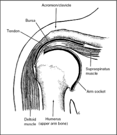
Description: Impingement of the shoulder, as the name denotes, refers to a compression  or pinching phenomenon of the soft tissues (rotator cuff tendons and subacromial bursa) between the arm bone or ball of the shoulder (humeral head) and the bone that forms the roof of the shoulder (acromion). It is a syndrome that is characterized by pain along the outer portion of the upper arm that worsens with overhead activities such as throwing, serving and or lifting. Patients often complain of strength loss associated with an aching pain that frequently will wake them at night. The rotator cuff muscle tendon units consist of four muscles that encapsulate the upper and outer portions of the ball of the shoulder (humeral head). Their function is to depress the humeral head in concert with the biceps tendon and stabilize the shoulder joint so that when the larger muscle groups fire (i.e. the deltoid) the arm is raised and the bones do not scrape against each other. Any time tendons cross a joint there is the increased risk of friction and tendon abrasion with use. In order to curtail this the body creates bursae, or fluid filled sacs, that engulf and protect the areas in jeopardy.
  The subacromial bursa of the shoulder sits over the top of the rotator cuff and allows for improved gliding of the cuff tendons between the shoulder bones with shoulder motion. If this bursa becomes inflamed acutely (suddenly) due to an injury or traumatic event; or if it is chronically irritated from a dysfunctional rotator cuff, bony prominences, improper lifting, throwing or job mechanics the space provided for these soft tissues becomes compromised and a downward spiral of increased swelling, pain and inflammation occurs. This is what is most frequently referred to as subacromial bursitis with rotator cuff tendonitis.
Historically the teaching was that impingement was the result of differently shaped bony roofs (acromions) of the shoulder, some of which inherently afforded less space for the soft tissues. These different shapes were classified as Type I, II and III with Type III having the least amount of available space (see figure). Patients with shoulder symptoms will frequently ask, “Is that bony spur or differently shaped acromion, the cause of all of my problems?” The answer is that it depends on their history and mechanism of injury. Sudden traumatic injuries in the face of a type III acromion can lead to impingement simply because the cuff tendons become pinched acutely and respond with inflammation and scarring.  On the other hand simply having a Type III acromion does not mean that you are destined to develop impingement and pain. The opposite is true as well where a person has lived their entire life without symptoms but have used their shoulder excessively for work and play. As the rotator cuff becomes degenerative its ability to keep the ball (humeral head) depressed and in the socket lessens. The result is a rotator cuff and bursa that can now be pinched by the bony roof above. If the roof is flat the pinching will be more evenly distributed and symptoms less likely or moderate in nature, if it is spiked or hooked then impingement symptoms to a greater degree are more likely to be present.
Risk Factors: Impingement is common in athletes of all ages and in middle-aged people based upon the activities being performed. Young athletes involved in contact sports such as football, wresting and weightlifting are at an increased risk. Additionally those athletes who use their arms overhead for swimming, baseball, and tennis are particularly vulnerable. Laborers who do repetitive lifting or overhead activities using the arm, such as paper hanging, construction, or painting are also susceptible. Pain may also develop as the result of minor trauma or spontaneously with no apparent cause. Additional risk factors include anything that causes the space available for the rotator cuff tendons and bursa to be compromised such as the different acromial types discussed above, the presence of calcium deposits or thickened tendons due to scarring.
Preventive Measures: Preventative measures are often neglected by laborers and athletes of all calibers. These measures include an appropriate warm up and stretch before work, practice or competition. Above all the use of proper technique may be the single most important factor when it comes to prevention.
General Treatment Considerations
Nonsurgical Treatment: Initial treatment consists of nonsteroidal anti-inflammatory medications and ice to relieve the pain. Physical therapy is typically prescribed focusing on stretching and strengthening exercises, and modification of the activity that initially caused the problem. Therapy may include a work station evaluation or technique modification teaching. If there is concern that a rotator cuff tear may also be present an MRI scan will usually be ordered. Subacromial injections of steroid and numbing medication may ultimately be ordered so long as a rotator cuff tear is not present. These injections reduce inflammation which can ultimately lead to long term pain relief. Care must be taken with activities immediately following an injection because they weaken muscle and tendon, hence making them more vulnerable to injury up to a period of three weeks post injection.
Surgical Treatment: When nonsurgical treatment does not relieve the pain or disability the Orthopaedic Surgeon may recommend surgery. The goal of surgery is to remove any bony projections or scarred bursa as well as address any other existing pathology (rotator cuff dysfunction) that may be contributing to the formation of impingement.
Operative Technique (What Is Done): Where as in the past large portions of the acromion were removed to afford greater space for the rotator cuff and bursa, recent literature has shown that a minimalist approach with the conversion of a hooked acromion (Type II or III) to simply a flat one (Type I) fairs significantly better with greater patient satisfaction and fewer complications. Different techniques are in use at this time but the overall goal remains the same; to decompress the subacromial space by removing the chronically inflamed bursa and release the thickened CA ligament while removing the acromial curve, hook, or bone spur. Arthroscopic techniques utilize a small camera and instruments that are inserted via small incisions, or portals, into the shoulder joint. This allows for other pathology within the joint itself to be addressed at the same sitting while maximizing visualization of the subacromial space. Electricity in addition to a motorized shaver is used to remove the bursa followed by a high speed burr for bony removal.
Rehabilitation: Postoperative rehabilitation and exercises are an integral part in achieving a successful surgical outcome. The ultimate goals are to decrease or eliminate the pain and to regain motion and strength so that a return to sport or activities of daily living can be initiated. The Orthopaedic surgeon will provide a rehabilitation program based on the patient’s needs and the findings at surgery. This will include exercises to regain range of motion of the shoulder and strength of the arm. Complete recovery typically can be expected around the 3 to 4 month postoperative period.
The Bottom Line
Anything that decreases the available space for the soft tissues (rotator cuff and bursa) so that they then become irritated and inflamed can lead to the formation of impingement or worsen it if already present. When attempting to diagnose impingement all potential causes and associated risk factors must be taken into consideration if a successful treatment outcome is sought.